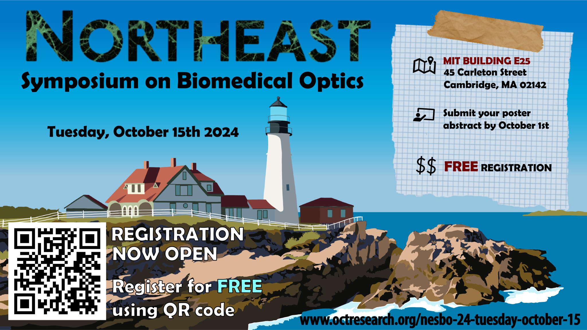
NESBO ’24 Tuesday October 15
About:
NESBO is an annual, one-day event aiming to bring together junior researchers from across the greater New England area to stimulate scientific discussion and promote collaboration within the local Biomedical Optics community. It is a great opportunity to interact with other Graduate students and Postdoctoral researchers in a relaxed atmosphere. We hope to see you there
Registration is closed.
Download the PDF program here (with posters list)
Schedule
Time | Event | Speaker | Affiliation |
|---|---|---|---|
| 08:30 | Coffee + Registration | ||
| 08:50 | Introduction | Prof. Brett E Bouma | Wellman Center for Photomedicine (MGH – HMS) |
Session 1: Advances in Contrast Mechanisms | |||
| 09:00 | Asynchronous, semi-reverberant elastography | Ginger Schmidt | Wellman Center for Photomedicine (MGH – HMS) |
| 09:30 | Exploring polarimetric imaging for label-free guidance of diagnostic and surgical procedures in oncology | Michael Singh | University of Toronto |
| 10:00 | Coffee Break | ||
Session 2: Fluorescence in Cancer | |||
| 10:20 | Pressure Enhanced Sensing of Tissue Oxygen: Guided surgical resection by detection of hypoxic tissue | Arthur Felix Petusseau | Optics in Medicine at Dartmouth Research Center |
| 10:50 | In Vivo Labeling and Detection of Circulating Tumor Cells with Fluorescent Molecular Contrast Agents | Josh Pace | Northeastern University |
| 11:20 | Panel discussion: Academia versus Industry |
|
|
| 12:20 | Lunch/Discussion | ||
Session 3: Computational Biophotonics | |||
| 13:20 | Towards streamlining the breast pathology workflow: automated processing of breast optical coherence tomography volumes | Arielle S. Joasil | Columbia University |
| 13:50 | NeuroPath: A Neuropathology-Specific Foundation Model for Advancing AI-Powered Analysis of Neurodegenerative Diseases | Shuying Li | Boston University |
| 14:20 | Developing medical diagnostics with computational biophotonics | Taylor L. Bobrow | Johns Hopkins University |
Keynote speaker | |||
| 14:50 | It Goes without Saying Taking the Guesswork Out of Your PhD in Engineering | Prof. Caroline Boudoux | Polytechnique Montréal |
| 15:30 | Break and Setup Posters | ||
| 15:40 | Poster Session | ||
| 16:55 | Concluding Remarks and Posters Awards | ||
| 17:15 | Social & Networking @ Cambridge Brewing Co. (17:30 – 19:30) | ||
Talks
Session 1: Advances in Contrast Mechanisms
 Asynchronous, semi-reverberant elastography
Ginger Schmidt, Wellman Center for Photomedicine (MGH-HMS)
Click to read abstract
Optical coherence elastography measures elasticity. Wave-based elastography leverages the properties of shear waves traveling through tissue primarily to infer shear modulus. These methods have seen significant development over the past decade; however, existing implementations in OCT require robust synchronization of shear wave excitation with imaging, complicating widespread clinical adoption. We present a method for complete recovery of the harmonic shear wave field in an asynchronous, conventional frame-rate, raster-scanning OCT system by modeling raster-scanning as an amplitude modulation of the displacement field. This technique recovers the entire spatially and temporally coherent complex-valued shear wave field from just two B-scans, while reducing the time scale for sensitivity to motion from minutes to tens of milliseconds. To the best of our knowledge, this work represents the first successful demonstration of reverberant elastography on a human subject in vivo with a conventional frame-rate, raster-scanning OCT system, greatly expanding opportunity for widespread translation.
Click to read speaker bio
Ginger Schmidt is graduate student researcher in the Center for Biomedical OCT Research and Translation (CBORT) within the Wellman Center for Photomedicine. She is studying Medical Engineering & Medical Physics through the Harvard-MIT Health Sciences and Technology (HST) PhD program. She is developing signal processing methods and hardware solutions to enable tissue elasticity measurements in vivo with elastography. Prior to her graduate studies, she studied engineering at Harvey Mudd College.
 Exploring polarimetric imaging for label-free guidance of diagnostic and surgical procedures in oncology
Michael Singh, University of Toronto
Click to read abstract
Polarimetry is emerging as a promising label-free cancer detection/imaging approach via its sensitivity to micro/macro structural changes during tumor development. Polarimetry currently lacks the specificity to yield diagnoses, however, it can be performed rapidly in a wide field-of-view. Thus it can instead be used to guide more sensitive but slower diagnostic tools to regions of interest thereby shortening their acquisition times. Our recent study employs wide-field polarimetric image guidance to target regions of interest on thick ex-vivo human breast to optimize mass spectrometry analysis. We also explore polarimetric contrast between cancerous and healthy tissue on the resected human breast specimens as well as on live pancreatic adenocarcinoma bearing mice.
Click to read speaker bio
Michael is a 5th year PhD student in Dr. Alex Vitkin’s laboratory as part of the Applied Biophotonics Division of Medical Biophysics at the University of Toronto. Michael completed his Bachelors degree in physics, and a Masters thesis on femtosecond laser-matter dynamics for molecular imaging and material ablation applications. Michael’s PhD studies focus on polarized light scattering dynamics in heterogenous media to advance the science of polarimetry into the biomedical domain. His recent research explores the use of polarimetric imaging as a potential tool to guide (1) mass spectrometric probing for optimized tissue diagnostics and (2) surgical resection of tumors.
Session 2: Fluorescence in Cancer
Pressure Enhanced Sensing of Tissue Oxygen: Guided surgical resection by detection of hypoxic tissue
Arthur Felix Petusseau, Optics in Medicine at Dartmouth Research Center
Click to read abstract
Fluorescent markers can make surgery more specific by enhancing contrast during tissue resection in certain types of disease. Pressure-Enhanced Sensing of Tissue Oxygenation (PRESTO) uses a commonly available human precursor molecule, 5-Aminolevulinic Acid, to stimulate immediate fluorescence when there is hypoxia present. This pre-contrast agent is metabolized into heme in most human cells, but produces a red fluorescent molecule, protoporphyrin IX, as an intermediate contrast agent. PpIX’s delayed fluorescence is amplified in low oxygen environment of tissue. PRESTO contrast can be used through tissue palpation, leading to contrast greater than 5 in pancreatic, brain, ovarian and head & neck tumors. PRESTO imaging is the first real-time widefield acquisition of palpation response in vivo, making it a valuable tool for highlighting hypoxic tissues and guiding oncologic surgical resection.
Click to read speaker bio
Arthur earned his Master’s degree in Photonics and Medical Engineering from the University of Strasbourg, France, in 2018, where he collaborated with Prof. Sylvain Gioux on developing digital holography methods for in vivo tissue imaging. He joined Brian Pogue’s lab at Dartmouth College in 2019, completing his Ph.D. in early 2023 with a focus on time-domain imaging of tissue metabolism. Currently, Arthur is a postdoctoral researcher in Prof. Bruza’s lab at Dartmouth College. His research interests include fluorescence imaging, oximetry, fluorescence-guided surgery, and FLASH radiation therapy.
In Vivo Labeling and Detection of Circulating Tumor Cells with Fluorescent Molecular Contrast Agents
Josh Pace, Northeastern University
Click to read abstract
Circulating tumor cells (CTCs) are the main facilitators of hematogenous metastasis. Our lab has developed a technique called ‘diffuse in vivo flow cytometry’ (DiFC) to non-invasively optically detect and enumerate CTCs from the tissue surface. Here, we report on combining DiFC with clinical stage fluorescent molecular contrast agents for the labeling and detection of CTCs in mice.
Click to read speaker bio
Josh Pace is a fifth-year graduate student pursing his Ph.D. in Bioengineering with a focus in biomedical optics at Northeastern University. Under the guidance of Mark Niedre, his research is focusing on the fluorescent detection of circulating tumor cells directly in the blood of mice. He received a B.Sc. Degree in Biomedical Engineering from the University of Arizona in 2019.
Session 3: Computational Biophotonics
Towards streamlining the breast pathology workflow: automated processing of breast optical coherence tomography volumes
Arielle S. Joasil, Columbia University
Click to read abstract
Breast cancer incidence is increasing yearly in the United States. This increased incidence necessitates the fast and accurate triaging of specimens for pathological examination. If data are acquired quickly, optical coherence tomography (OCT) has the potential to impact breast pathology. Recovering undersampled volumes with compressed sensing (CS) can allow for faster acquisition. In this talk, I will discuss the CS reconstruction of human breast OCT volumes and the diagnostic accuracy of these volumes.
Click to read speaker bio
Arielle S. Joasil is a fourth-year PhD candidate in the Electrical Engineering Department at Columbia University. She works with Dr. Christine P. Hendon in the Structure Function Imaging Laboratory, where she applies machine learning and image processing techniques to detect disease in optical coherence tomography volumes of the breast and aid the breast pathology process.
NeuroPath: A Neuropathology-Specific Foundation Model for Advancing AI-Powered Analysis of Neurodegenerative Diseases
Shuying Li, Boston University
Click to read abstract
Conventional neuropathological evaluations, whether for research or clinical diagnosis, are time-intensive and heavily reliant on expert interpretation, often leading to significant variability in results. This subjectivity underscores the need for more precise and efficient approaches. Here, we introduce NeuroPath, the first foundation model designed specifically for neuropathology. By leveraging digital neuropathology WSI database at the Boston University Alzheimer’s Disease Research Center (ADRC), NeuroPath aims to transform the analysis of diseases such as Alzheimer’s disease (AD), chronic traumatic encephalopathy (CTE), and other NDs. NeuroPath is developed using DINOv2 self-supervised learning (SSL) techniques and its performance is further evaluated through slide-level neuropathological tasks using attention-based multiple instance learning, including predictions of CTE staging. Our approach offers enhanced accuracy and reliability for analyzing complex neuropathological data and can potentially transform the field of digital neuropathology, facilitating faster, more accurate, and predictive assessments that could revolutionize our understanding and treatment of NDs.
Click to read speaker bio
Shuying Li is a Postdoctoral Associate in the Department of Electrical & Computer Engineering at Boston University. She earned her Ph.D. in Biomedical Engineering from Washington University in St. Louis. Her research integrates artificial intelligence (AI) with biophotonics, focusing on leveraging advanced AI tools to enhance optical imaging techniques for improved diagnostic precision and deeper insights into disease mechanisms. In her current role, she applies AI algorithms to human brain images captured by Polarization-Sensitive Optical Coherence Tomography (PS-OCT), Quantitative Birefringence Microscopy (qBRM), and neuropathological slides, striving to unlock new dimensions of neurodegenerative disease understanding.
Developing medical diagnostics with computational biophotonics
Taylor L. Bobrow, Johns Hopkins University
Click to read abstract
Computational biophotonics combines optical imaging systems and software algorithms to measure quantitative, diagnostic information from imaged tissue. I will present our research in developing and translating computational biophotonics technologies for a variety of important healthcare needs, including (1) preventing colorectal cancer with a computational colonoscope and deep learning, (2) detecting cather-associated urinary tract infections with lens-free holographic imaging, and (3) increasing access to eye care in low-resource settings with a low-cost, handheld wavefront aberrometer.
Click to read speaker bio
Taylor Bobrow is a Malone Postdoctoral Fellow in the Department of Biomedical Engineering at the Johns Hopkins University. He completed his BS in electrical engineering at Old Dominion University in 2017 and received his PhD in biomedical engineering from the Johns Hopkins School of Medicine in 2024. His current research is focused on using computational imaging to enable low-cost, continuous point-of-care monitoring with applications such as the prevention of hospital-acquired infections.
Keynote Speaker
It Goes without Saying
Taking the Guesswork Out of Your PhD in Engineering
Prof. Caroline Boudoux, Polytechnique Montréal
Click to read abstract
The definitive toolkit for doctoral students in engineering on thesis—and journal article—preparation, project (and stress) management, IP protection, collaborations, and other aspects of the PhD journey.
It shouldn’t take a PhD to get a PhD, but sometimes the process can seem that confusing—even though, to the mentors and advisors, so obvious that it goes without saying. For doctoral students in engineering confronting this dilemma, Caroline Boudoux, an accomplished researcher and entrepreneur, provides a demystifying guide to the challenges—daunting, seemingly routine, and at times unexpected—of pursuing a PhD in this demanding field. In It Goes without Saying, Boudoux marshals her considerable experience mentoring graduate students, teaching doctoral workshops, and—not so long ago—earning her own PhD at MIT to give PhD candidates the know-how, and the confidence, to succeed.
Click to read speaker bio
Caroline Boudoux is Professor of Engineering Physics at Polytechnique Montréal and Cofounder and Copresident at Castor Optics, a company commercializing a new line of double-clad fibers couplers. She is on the Board of Meetings for OPTICA, is a Fellow of SPIE, and a Fulbright scholar. Prof. Boudoux obtained her PhD from the Harvard-MIT Health Sciences and Technology program (USA) and completed a post-doctoral fellowship at Ecole Polytechnique (France) before starting her laboratory, in 2007, at Polytechnique Montreal (Canada). Her research topics range from laser-tissue interactions to novel instruments for biomedical imaging.
Panel discussion: Academia versus Industry
 Yong-Chul Yoon recently received his PhD from the Health Science and Technology (HST) at MIT, where he developed a novel optical coherence tomography (OCT) platform for identifying nerves during surgery under the supervision of Benjamin J. Vakoc at the Massachusetts General Hospital. Currently, he is finishing up his research as a post doc in the same lab while co-founding a start-up company that seeks to tackle the extremely difficult challenge of non-invasively inferring glucose concentration in vivo. Outside of research, he enjoys drumming for the Midnight Motion, a Boston-based funk R&B band.
 Nili Persits, the founder and CEO of Dottir Labs, has a Ph.D. in electrical engineering from MIT and over 10 years of industry experience of sensor design and integration. Her technology focuses on the scaling of optical spectroscopic Raman sensors to enhance sustainability across multiple industries including chemical manufacturing, food & beverage, agriculture, and environmental monitoring. Nili is a 2023 Activate fellowship recipient and has also received numerous grants and awards supporting women in STEM and entrepreneurship.
 Christa Haase is an Assistant Professor of Bioengineering and Physics at Northeastern University. Her lab focuses on uncovering how microenvironmental cells impact fate decisions in blood stem cells and drive the development and recurrence of blood cancers, such as leukemia. To achieve this, they design and apply cutting-edge spatial, single-cell, and optical technologies that enable the analysis of microenvironmental cells in vivo. Prior to joining Northeastern University, she was an Instructor at the Wellman Center for Photomedicine at Harvard Medical School and worked with the laboratory of Dr. Charles Lin, where she developed Image-seq, a technology that provides single-cell transcriptional and multi-omics data on cells that are isolated from specific spatial locations under image guidance. The technique is compatible with intravital microscopy and makes it possible to integrate the spatial, temporal and molecular information of the target cells. Moreover, it is uniquely suited to study rare cell populations in bone marrow tissue, including hematopoietic stem cells, leukemia initiating cells and stromal cells.
 David Veysset earned his PhD in Physical Chemistry from MIT in 2016 before completing postdoctoral training at MIT and Stanford University. In 2022, he co-founded Aluminio, a climate tech start-up, in 2022 and was a Breakthrough Energy Fellow. In 2024, he joined the Wellman Center for Photomedicine at MGH as a Physicist and Instructor. His current research focuses on developing novel optical diagnostics tools, bridging optics, acoustics, and spectroscopy.
 Sarat Chandra Gundavarapu is the Director of Photonics and Reader Development at SiPhox Health. He has over 10 years of experience in varied fields of integrated photonics including low-noise lasers, biomedical imaging, and optical sensors. Sarat received his Ph.D. at University of California, Santa Barbara developing silicon nitride integrated photonic Brillouin lasers and was a research fellow at Harvard Medical School and Massachusetts General Hospital, developing portable optical systems for clinical and translational imaging.