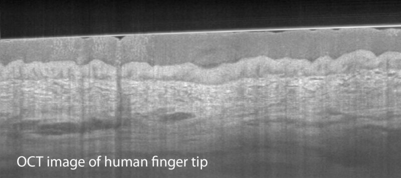
Optical coherence tomography (OCT) is an imaging modality that uses near infrared light to provide cross-sectional views of the subsurface microstructure of biological tissue. Similar to ultrasound imaging, it measures the delay of the signal reflected from different depths within the sample, but using light and interferometric detection instead of sound. This provides high spatial resolution of ~10 µm. Volumetric images are obtained by scanning the OCT beam across the sample surface. The light is scattered and reflected by abrupt changes in the refractive index of the tissue, generating intrinsic contrast without the need for any labeling. Scattering, however, also scrambles the forward propagating beam and limits the imaging depth to 1-2 mm.
OCT has evolved into a powerful microscopy technique for a broad range of applications in biology and clinical medicine. OCT can readily be performed through flexible, narrow diameter optical fiber probes for imaging internal organs. Recent advances, such as optical frequency domain imaging (OFDI), have increased OCT imaging speeds by nearly two orders of magnitude, enabling microscopic, three-dimensional imaging over large fields-of-view, a capability that cannot be provided by conventional optical imaging or histology. Today, OCT is commercially available and routinely used for imaging of the retina and the skin, and, implemented into a catheter, for imaging of the coronary arteries and the upper gastrointestinal tract.