Quantitative Microstructure Sensing
Contrast in optical coherence tomography (OCT) results from light scattering events in tissue. The behavior of these events is governed by the scattering-based optical properties of imaged tissue samples, which in turn are governed by the physical characteristics of the tissue on a subcellular scale. By calculating optical properties from the OCT intensity signal, it is possible to extract structural information about an imaged sample related to features far below the typical resolution of a clinical OCT system. Physical sample characteristics calculable using these methods include the sizes and concentrations of scattering particles, such as cell organelles. The ability to volumetrically extract this information from biological samples is attractive for a number of clinical applications, such as the early detection of dysplasia through sensitivity to precancerous enlargement of cell nuclei and hypercellularity.
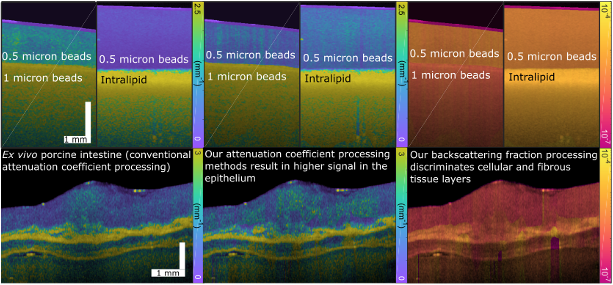
Our approach to microstructure sensing focuses on the determination of the attenuation coefficient, an analogue at typical OCT wavelengths of the scattering coefficient, and the backscattering fraction; each of these optical properties is each sensitive to the sizes, refractive indices, and shapes of scattering particles in tissue, and the attenuation coefficient further adds sensitivity to particle concentration. We are developing methods to quantify each of these parameters accurately and volumetrically for diverse tissue samples. Our attenuation coefficient calculation methods improve on previous approaches and enable the computation of a layer-resolved backscattering fraction for the first time in tissue. The complementary information offered by these two optical properties reduces the ambiguity in interpreting the physical meaning of the scattering signal, and progresses towards precise measurements of the sizes and concentrations of scattering particles, such as nuclei, in tissue. We are further expanding our methods to account for complicating phenomena such as multiple scattering and the behavior of cylindrical scatterers (such as extracellular matrix components or collagen fibers) in the effort to make our analysis fully quantitatively translatable to broader biomedical imaging applications.
Key Researchers
Relevant Publications
- Taylor M. Cannon, Brett E. Bouma and Néstor Uribe-Patarroyo, “Layer-based, depth-resolved computation of attenuation coefficients and backscattering fractions in tissue using optical coherence tomography.” Biomed. Opt. Express 12, 5037-5056 (2021).
- Taylor M. Cannon, Brett E. Bouma and Néstor Uribe-Patarroyo, “Mapping optical scattering properties to physical particle information in singly and multiply scattering samples.” Biomed. Opt. Express 14, 4326–4348 (2023).