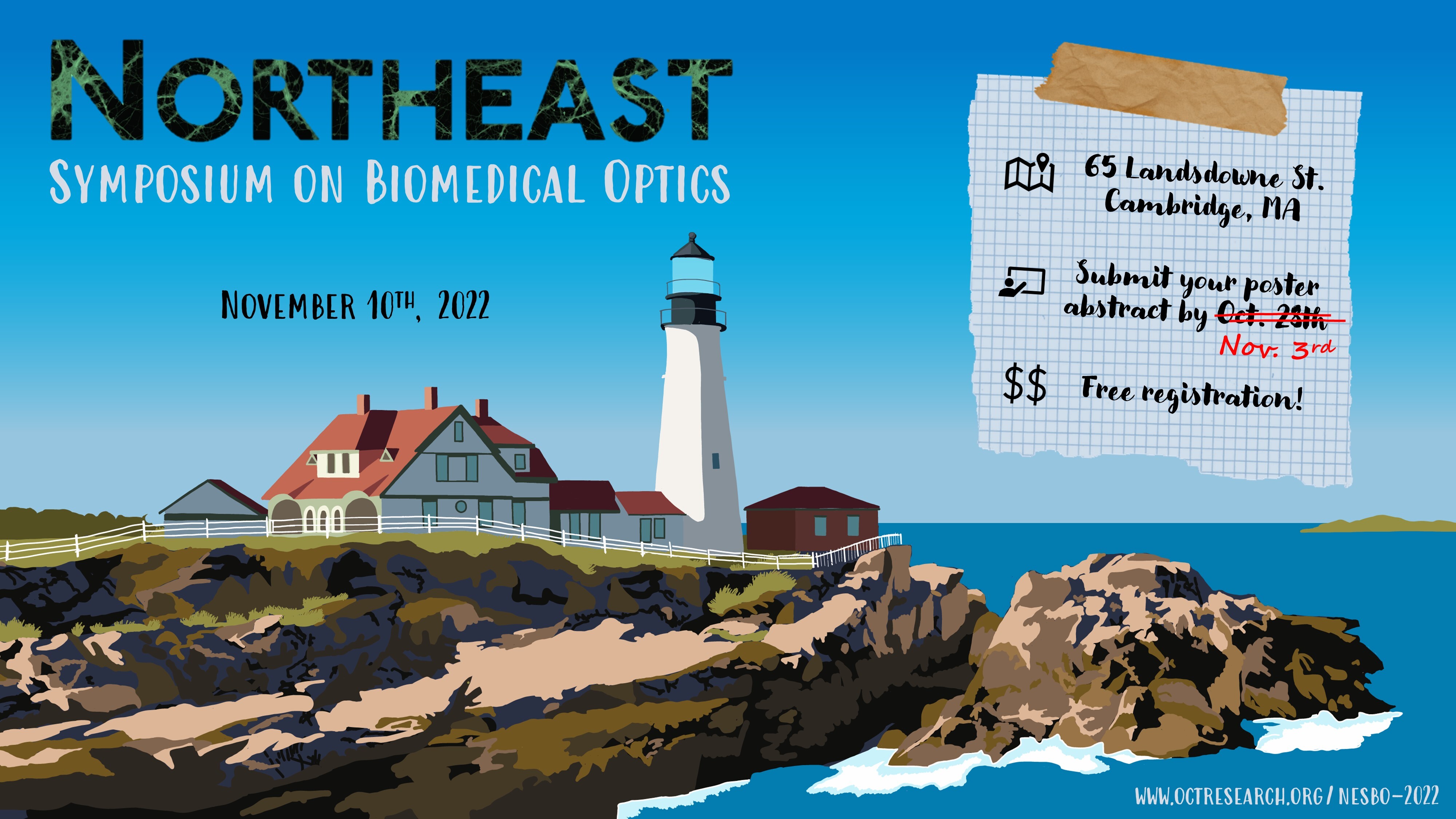
NESBO ’22 Thursday November 10
About:
NESBO is an annual event aiming to bring together junior researchers from across the greater New England area to stimulate scientific discussion and promote collaboration within the local Biomedical Optics community. It is a great opportunity to interact with other young researchers in a relaxed atmosphere. We hope to see you there!
Registration is now closed.
Dear poster presenters: please limit the size of the poster to no larger than 48″ X 36″, landscape orientation.
Schedule
Time | Event | Speaker | Affiliation |
|---|---|---|---|
| 08:30 | Coffee + Badge Distribution | ||
| 09:00 | Introduction | Brett E. Bouma | MGH-Wellman |
Session 1: Deep Learning in Biomedical Optics | |||
| 09:15: | Computational Miniature Mesoscope with a fast and reliable deep learning reconstruction framework | Qianwan Yang | Boston University |
| 09:40 | Deep learning for detection of breast disease in optical coherence tomography | Diana Mojahed | MIT |
| 10:05 | Coffee Break | ||
Session 2: Neurophotonics | |||
| 10:30 | Kilohertz two-photon microscopy for high-throughput deep tissue imaging of fast biology dynamics | Sheng Xiao | Boston University |
| 10:55 | All-optical electrophysiology reveals mechanisms of dendritic integration in vivo. | David Wong-Campos | Harvard University |
| 11:20 | Panel: Academia to Industry |
|
|
| 12:30 | Lunch/Discussion | ||
Session 3: Advances in Imaging Methods and Optical Design | |||
| 13:15 | Mapping cellular and molecular architecture of mammalian cortical evolution by single cell transcriptome-scale imaging | Rongxin Fang | Harvard University |
| 13:40 | Towards realizable optical meta-surfaces through physics-informed quantization aware training | Ramith Hettiarachchi | Harvard University |
| 14:05 | A multi-beam, circular-ranging OCT system for 3D, video-rate, laparoscopic imaging | Yongjoo Kim | MGH-Wellman |
| 14:30 | Coffee Break | ||
| 15:00 | Poster Session | ||
| 16:30 | Concluding Remarks and Awards | ||
| 17:00 | Social Hour @ Cambridge Brewing Co.(17:00 – 19:00, click for location) | ||
Invited Talks
Deep Learning in Biomedical Optics
Computational Miniature Mesoscope with a fast and reliable deep learning reconstruction framework
Qianwan Yang, Boston University
Click to read abstract
Computational Miniature Mesoscope (CM2) is a novel fluorescence imaging device that achieves single-shot large-scale volumetric imaging on a compact platform by jointly designing the optics and algorithm. Here, we demonstrate an advanced CM2 system with a deep learning reconstruction framework, termed CM2Net, to perform high resolution 3D reconstruction with fast speed. The CM2Net is fueled by simulated realistic field varying data generated from a 3D-LSV forward model and is experimentally validated by imaging phantom objects with embedded fluorescent particles. We demonstrate that the CM2Net achieves ~8X better axial localization and ~1400X faster reconstruction speed as compared to model-based deconvolution.
Click to read speaker bio
Qianwan Yang is currently a third year Ph.D. student working with Professor Lei Tian in the Computational Imaging Systems Lab at Boston University. Her research interests center around computational imaging, with particular emphasis on developing novel Computational Miniature Mesoscope for large scale 3D fluorescence imaging.
 Deep learning for detection of breast disease in optical coherence tomography (OCT) images
Diana Mojahed, Ph.D. MIT
Click to read abstract
Technologies allowing for real-time histology evaluation may transform the entire anatomic diagnostic process across multiple organs – from biopsy to intraoperative margin assessment to tissue grossing as well as tissue banking. Deep learning can greatly improve the speed and efficiency with which optical coherence tomography (OCT) images are ready in this context. We show our work towards developing artificial intelligence (AI) for cancer detection within OCT images. We developed a convolutional neural network (CNN) to classify OCT images of breast tissue from 49 patients. The binary cancer classification achieved 94% accuracy, 96% sensitivity, and 92% specificity. This framework had higher accuracy than the 88% accuracy of 7 clinician readers combined from a multi-reader study.
Click to read speaker bio
Diana Mojahed is a Postdoctoral Associate in Materials Science & Engineering at MIT. She graduated with her PhD in Biomedical Engineering in 2021 from Columbia University working under Dr. Christine Hendon. In her PhD, she developed an optical coherence tomography (OCT) to rapidly evaluate breast biopsies and pathology specimens to improve the quality of care for patients. Her device and artificial intelligence (AI) algorithms were demonstrated in a pilot 100-patient clinical study. Her work was supported by the Columbia BiomedX Technology Accelerator Program, which is a highly competitive program that choses 5-6 projects per year to have strong potential for advancing clinical practice.
Neurophotonics
 Kilohertz two-photon microscopy for high-throughput deep tissue imaging of fast biology dynamics
Sheng Xiao, Ph.D. Boston University
Click to read abstract
High-speed optical microscopy is essential for the observing fast biological events. We present a kilohertz frame rate two-photon microscope based on a recently developed device called scan multiplier unit. Our technique enables a conventional video-rate two-photon microscope to be converted for kilohertz frame rate imaging, with no compromise in excitation efficiency or penetration depth. We demonstrate our system on in vivo imaging of neurovascular dynamics over a field-of-view of 200×200 um, and penetration depth > 500 um.
Click to read speaker bio
Dr. Xiao received his B.S. in Optical Engineering from Huazhong University of Science and Technology, and Ph.D. in Electrical Engineering from Boston University. He is currently a research scientist at the Biomicroscopy lab at Boston University. His research interests include the development of novel optical imaging and microscopy techniques and their applications in neuroscience studies.
All-optical electrophysiology reveals mechanisms of dendritic integration in vivo.
David Wong-Campos, Ph.D. Harvard University
Click to read abstract
Neurons convert synaptic inputs to spiking outputs, yet we lack a coherent picture of how dendrites integrate synaptic inputs to produce spikes, and how back-propagating spikes (bAPs) interact with the dendritic arbor. The ability to map voltage dynamics at high resolution throughout a dendritic tree would be a transformative capability for studying how individual neurons perform computations. Here, we combine patterned channelrhodopsin activation and structured one-photon voltage imaging, to control and measure electrical activity throughout a dendritic tree in vivo. We estimate electrical cross-couplings among dendritic branches and measure failures of bAPs, which may have consequences for brain plasticity.
Click to read speaker bio
David Wong-Campos was born in Chiclayo, Peru and attended the Monterrey Institute of Technology and Higher Education (ITESM) in Mexico for his undergraduate. He then completed his Ph. D. in Physics at University of Maryland under the direction of Christopher Monroe, where he implemented a novel quantum logic gate and performed high resolution imaging experiments on individual atoms. He took a two-year industry job at a quantum computing startup (IonQ) and held an advisory position at a cancer company (Delee). He returned to academia and currently pursues postdoctoral training with Adam Cohen in high resolution in vivo voltage imaging.
Advances in Imaging Methods and Optical Design
 Mapping cellular and molecular architecture of mammalian cortical evolution by single cell transcriptome-scale imaging
Rongxin Fang, Ph. D. Harvard University
Click to read abstract
The human cerebral cortex has tremendous cellular diversity. How different cell types are organized in the human cortex and how cellular organization varies across species remain unclear. We performed spatially resolved single-cell profiling of 4000 genes using multiplexed error-robust fluorescence in situ hybridization (MERFISH), generated a molecularly defined and spatially resolved cell atlas of the human cortices. We further explored cell-cell interactions arising from soma contact or proximity in a cell type–specific manner. Comparison of the human and mouse cortices showed conservation in the laminar organization of cells and differences in somatic interactions across species. Our data revealed human-specific cell-cell proximity patterns and a markedly increased enrichment for interactions between neurons and non-neuronal cells in the human cortex.
Click to read speaker bio
Dr. Rongxin Fang is a postdoctoral fellow at Harvard University and is a recipient of the HHMI-Damon Runyon postdoctoral fellowship. He received his Ph.D. from UC San Diego in 2019. During his PhD, he was advised by Dr. Bing Ren and developed sequencing-based techniques to perform high-throughput measurements of enhancer activity, function to structure, and further applied these techniques to understand how enhancers contribute to the cellular diversity in the brain. As a postdoctoral fellow, he is currently working with Dr. Xiaowei Zhuang, Dr. Adam E. Cohen and Dr. Bruce Yankner and developing optic techniques to image the molecular and cellular organization of the mammalian brain, to understand how spatial organization changes during evolution and how communications between different cell types contribute to neuronal function in healthy and diseased brain.
 Towards realizable optical meta-surfaces through physics-informed quantization aware training
Ramith Hettiarachchi, Post Baccalaureate Fellow, Harvard University
Click to read abstract
Computational imaging performs intelligent measurements with a “brain” made of programmed optics like meta-surfaces. These programmable optics – though packed with billions of linear operations in a cubic millimeter – often performs poorly due to fabrication constraints. Here we propose a physics-informed quantization-aware training framework that accounts for these constraints and achieves robust designs. We discuss two types of metasurfaces, a learnable Fourier filter, and a diffractive deep neural network for applications such as phase imaging and phase object classification while accounting for the aforementioned fabrication constraints.
Click to read speaker bio
Ramith is a Joint Post-Bac Fellow affiliated to Wadduwage Lab and So Lab in the Division of Science at Harvard University. He completed his B.Sc. degree from University of Moratuwa, Sri Lanka in Electronic Engineering. As a Post-Bac Fellow at Wadduwage Lab, he is currently working with Dr. Dushan Wadduwage on making learnable optical systems robust and realizable by factoring in practical considerations such as fabrication constraints. His research interests include using machine learning for scientific discovery and focusing on the robustness, interpretability, and equitability of machine learning algorithms.
A multi-beam, circular-ranging OCT system for 3D, video-rate, laparoscopic imaging
Yongjoo Kim, Ph. D. MGH-Wellman
Click to read abstract
A 32-channel circular-ranging (CR)-OCT system was developed to facilitate the data-efficient acquisition of volumetric OCT data in surgical fields. A high-speed laser source and beam parallelization have increased the A-line rate to over 10 MHz. The novel system design incorporating a frequency comb source, a high-power optical amplifier, an all-fiber-based, 32-channel interferometer, a 32-beam scanning laparoscope, and 32-channel detection channels will be presented. We have demonstrated real-time volumetric CR-OCT imaging of a patterned sample which allowed the registration of the images from each channel to form a merged image sequence and a tissue sample. We also performed polarization-sensitive imaging on muscle and nerve using a custom-designed polarization modulation circuit devised for polarization-diverse detection in the multi-beam approach.
Click to read speaker bio
Yongjoo Kim is a Research Fellow at Harvard Medical School and the Wellman Center for Photomedicine. He received his PhD in Mechanical Engineering at Korea Advanced Institute of Science and Technology (KAIST). His research interest is multi-beam, high-speed circular-ranging OCT imaging.
Academia to Industry Panel
Mohammad Haft-Javaherian is the machine learning manager at Iterative Health, whose mission is to develop technology that can benefit patients anywhere, regardless of geographic or socioeconomic barriers. He was Bullock Postdoctoral Research Fellow at Wellman Center HMS and MIT CSAIL, working with Prof. Brett Bouma and Prof. Polina Golland in close collaboration with Prof. Martin Villiger. He received his Ph.D. in Biomedical engineering from Cornell University, working with Prof. Nozomi Nishimura and Prof. Chris Schaffer and collaborating closely with Mert Sabuncu. His research interest centered on developing computer vision and machine learning techniques for biomedical image analysis.
Ethan LaRochelle received his PhD in 2020 from the Thayer School of Engineering at Dartmouth College where he was advised by Brian Pogue and Kim Samkoe. Ethan’s PhD research focused on the clinical translation of in vivo fluorescent and phosophorescent imaging for applications at the intersection of photodynamic and radiation therapies. He is a 2017 recipient of the National Science Foundation GRFP Fellowship. In 2019 he co-founded QUEL (Pronunciation: /kɛl/) Imaging with Alberto Ruiz and Brian Pogue, and is currently the CEO. Ethan has been the PI for two SBIR Phase I awards and is currently the PI of an active NIH-NIBIB SBIR Phase II grant. Ethan is originally from Maine and currently lives in a log house in Vermont.
Dr. Michael J. Williams is from Philadelphia, PA and a recent 2018 Ph.D. graduate from Delaware State University in Optics. He received his bachelors of science degree in Physics in 2009 from Morehouse College in Atlanta, GA and he received his master’s degree in materials science in 2012 from Fisk University in Nashville, TN. His Ph.D. research was the investigation of the linear, nonlinear, and fluorescent characterization of various nanodiamond suspensions using well-established characterization methods and techniques. The purpose was to determine a deeper understanding on how to engineer nanodiamonds to enhance their optical properties for lasers, biophotonics, and quantum optical applications. Currently he is an Applications Scientist for Boston Electronics, a photonics distribution company who develop and manufacture superior advanced electro-optical products, and perform leading edge research by providing customers with a broad range of solutions and knowledgeable application support. He is also serves as an OPTICA Ambassador, part of a community of emerging leaders in the optics and photonics community.
Samantha Paulsen has a background in cell therapy and tissue engineering. As a graduate student at Rice University, she is focused on using 3D printing and fluid dynamics modeling to develop in vitro and in silico models for blood flow in complex vascular architectures. After completing her PhD at Rice University, she worked for 2 years as a project engineer in bioprocessing at GE Healthcare, now Cytiva. For the past two years she has been working in drug product development for cell therapies with the Astellas Institute for Regenerative Medicine. In her role, she is responsible for developing new methods to cryopreserve cells and deliver cell therapies to patients in the clinic.
Sheldon J.J. Kwok, Ph.D. is CEO and co-founder of LASE Innovation, a startup spin-off from Prof. Seok-Hyun Yun’s lab at Massachusetts General Hospital. Dr. Kwok received his bachelor’s degree in Chemical Physics at Columbia University in New York City. Afterwards, he enrolled in the MD/PhD program at Harvard and MIT. Dr. Kwok received his PhD in Medical Engineering and Medical Physics at MIT, completing his thesis work in Prof. Yun’s lab in 2019. His research on the development of novel laser particle technology for single-cell tracking is being commercialized by LASE Innovation for life science R&D markets. The company has raised over $7 MM in equity financing and currently has 12 employees headquartered in Woburn, MA.
NESBO 2022 Executive Committee:
- Pelham Keahey, PhD — contact
- Danielle J. Harper, PhD — contact
- Jian Ren, PhD — contact
- Yong-Chul Yoon, BS/BA — contact