Glaucoma is a leading cause of irreversible blindness with sixty million people affected worldwide. Better retinal imaging methods hold the potential for improved early detection, since structural tissue damage often precedes vision loss. The most commonly used glaucoma imaging parameter in spectral-domain OCT instruments is retinal nerve fiber layer thickness for which thinning is a strong indicator of glaucoma. Current evaluation of glaucomatous nerve fiber layer changes relies on the thickness or the area of the defects, but little has been reported on three-dimensional volumetric changes. The current project is the first to directly calculate peripapillary 3D retinal nerve fiber layer volume and to evaluate its diagnostic capability in both normal and glaucoma patients in a multiethnic United States population. Custom-built software was developed to determine the 3D retinal nerve fiber layer volume from commercial spectral-domain optical coherence tomography scans. The thickness of several retinal layers was analyzed with image segmentation and integrated over annular retinal quadrant areas around the optic disc for volumetric measurements. Among all annular quadrants the best diagnostic capability was associated with the inferior quadrant. Otherwise global 3D volumetric measurements were comparable to global 2D thickness results. Overall 3D retinal nerve fiber layer volume showed excellent diagnostic performance for detecting glaucoma.
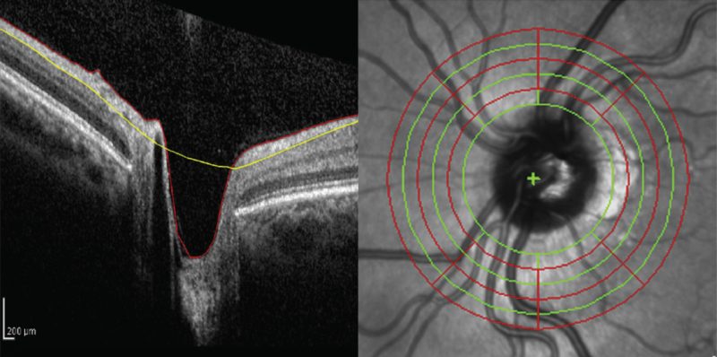
In the figure on the left one of the B-scans of the 3D volume set is shown, which was automatically segmented using edge and intensity information. On the right a depiction of how the software defines 4 annuli for volumetric analysis by superimposing 6 circles on the infrared reflectance image of the 3D volume scans is shown.
The Center for Biomedical OCT Research and Translation provided support in the development of the custom software for the analysis of the spectral-domain OCT datasets.
Key Researchers
- Benjamin J. Vakoc
- Brett E. Bouma
- Teresa C. Chen
- Boy Braaf
Relevant Publications
- Ziad Khoueir, Firas Jassim, Linda Yi-Chieh Poon, Edem Tsikata, Geulah S. Ben-David, Yingna Liu, Eric Shieh, Ramon Lee, Rong Guo, Georgia Papadogeorgou, Boy Braaf, Huseyin Simavli, Christian Que, Benjamin J. Vakoc, Brett E. Bouma, Johannes F. de Boer, Teresa C. Chen, Diagnostic Capability of Peripapillary Three-dimensional Retinal Nerve Fiber Layer Volume for Glaucoma Using Optical Coherence Tomography Volume Scans. American Journal of Ophthalmology 182, 180–193 (2017).