Percutaneous and Interstitial Imaging
The goal of this TRD project is to enhance the power and functionality of endoscopic and probe-based OCT. OCT’s compact and flexible form factor has enabled its integration into miniature probes, catheters, interstitial needles, micro-endoscopes, and compact hand-held devices. It affords the capacity to reach remote organs of the human body, enabling OCT to be routinely used for clinical investigation of the coronary arteries, the gastrointestinal tract, and the lung. This TRD is developing instruments and algorithms to overcome fundamental barriers, intrinsic to flexible, rotational fiber-optic probes, that have previously compromised image quality and prohibited the integration of flow measurements and polarization-metrics into this imaging geometry.
Flow imaging
Blood flow is the single most important attribute of arterial function. Stenotic atherosclerotic lesions in the coronary arteries become clinically significant when they impair the blood supply distal to the lesion and critically limit oxygen supply to the myocardium. To avoid the use of multiple intravascular catheters, we are working towards deriving blood flow measurements from OCT imaging with a standard intravascular imaging catheter. This would simplify the procedure and provide a functional assessment of lesion significance together with the detailed structural plaque features used to guide the intervention. To overcome limitations due to phase instability and accurately measure the mostly lateral flow surrounding the catheter, we are refining strategies that deduce flow from correlation-based measurements. Specific challenges arise from the presence of flow gradients within the coherence volume, as well as multiple forward scattering induced by red blood cells in blood.
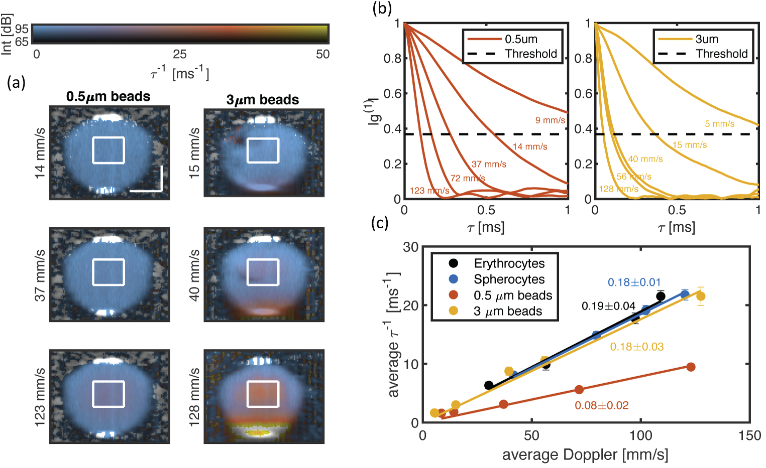 Decorrelation signal of 3µm and 0.5µm polysterene beads in a flow phantom. Comparison with Doppler signal in controlled bench-top setting reveals the increased decorrelation signal induced by multiple forward scattering in the 3µm beads (adapted from Ferris et al. Biome. Opt. Express 11, 1947-1966 (2020).)
Polarization imaging
Polarization-sensitive optical coherence tomography (PS-OCT) measures the polarization state of the light scattered as a function of its path length travelled into tissue. Many tissues with a fibrillar architecture exhibit birefringence, which results in a path length-dependent variation of the polarization state. PS-OCT can provide insight into the make-up and physical orientation of this tissue microstructure on a scale beyond the imaging resolution of OCT and thereby afford contrast between tissues that are indiscernible in OCT’s conventional scattering signal. Fiber and catheter-based polarization imaging requires compensating for the polarization effect of system-induced polarization distortions, catheter motion, and preceding tissue layers. Applied to imaging of the coronary arteries in human patients, intravascular polarimetry reveals birefringence of collagen and smooth muscle cells in the coronary arterial wall, while the presence of lipid and macrophages cause depolarization, offering critical insights into important features of coronary atherosclerosis. The same contrast mechanism also hold promise for imaging of white matter tracts in the brain.
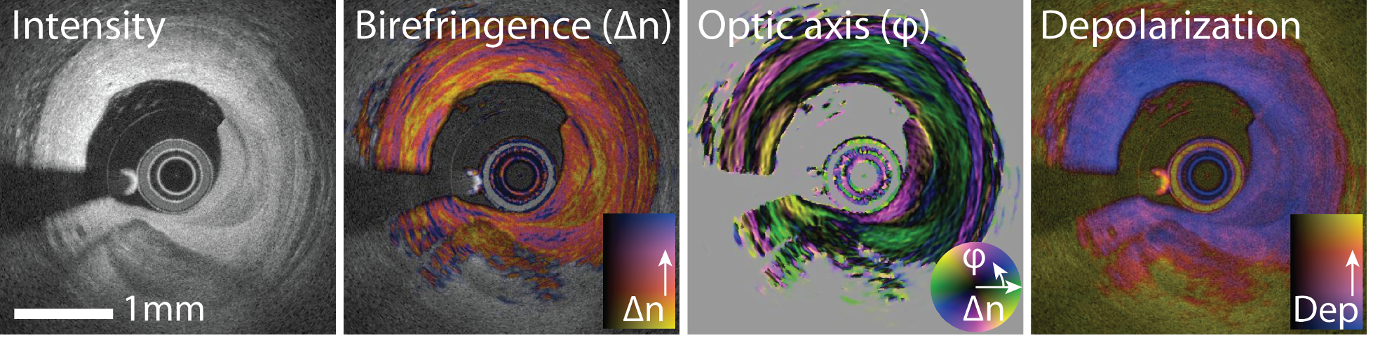 Intravascular polarimetry in the right coronary artery of a 69-year-old man, complementing the scattering intensity tomogram with quantitative measurements of depth-resolved tissue birefringence and optic axis orientation, in addition to depolarization.
Miniature rotation probes
We are developing miniature rotational, side-looking fiber probes with a diameter <300µm. The short catheters are interfaced through a catheter-connector with a sturdy tether, consisting of an inner drive shaft comprising the fiber encapsulated in a static sleeve. The tether transmits the torque from the proximal motor drive unit to the probe. The tether is connected to the catheter with standard angle-polished fiber connectors and a mating sleeve held by bearings and encapsulated in a cylindrical casing. This body fits inside a 10mL syringe, which conveniently can serve as pullback unit, actuated by a linear stage for synchronized pullback measurements.
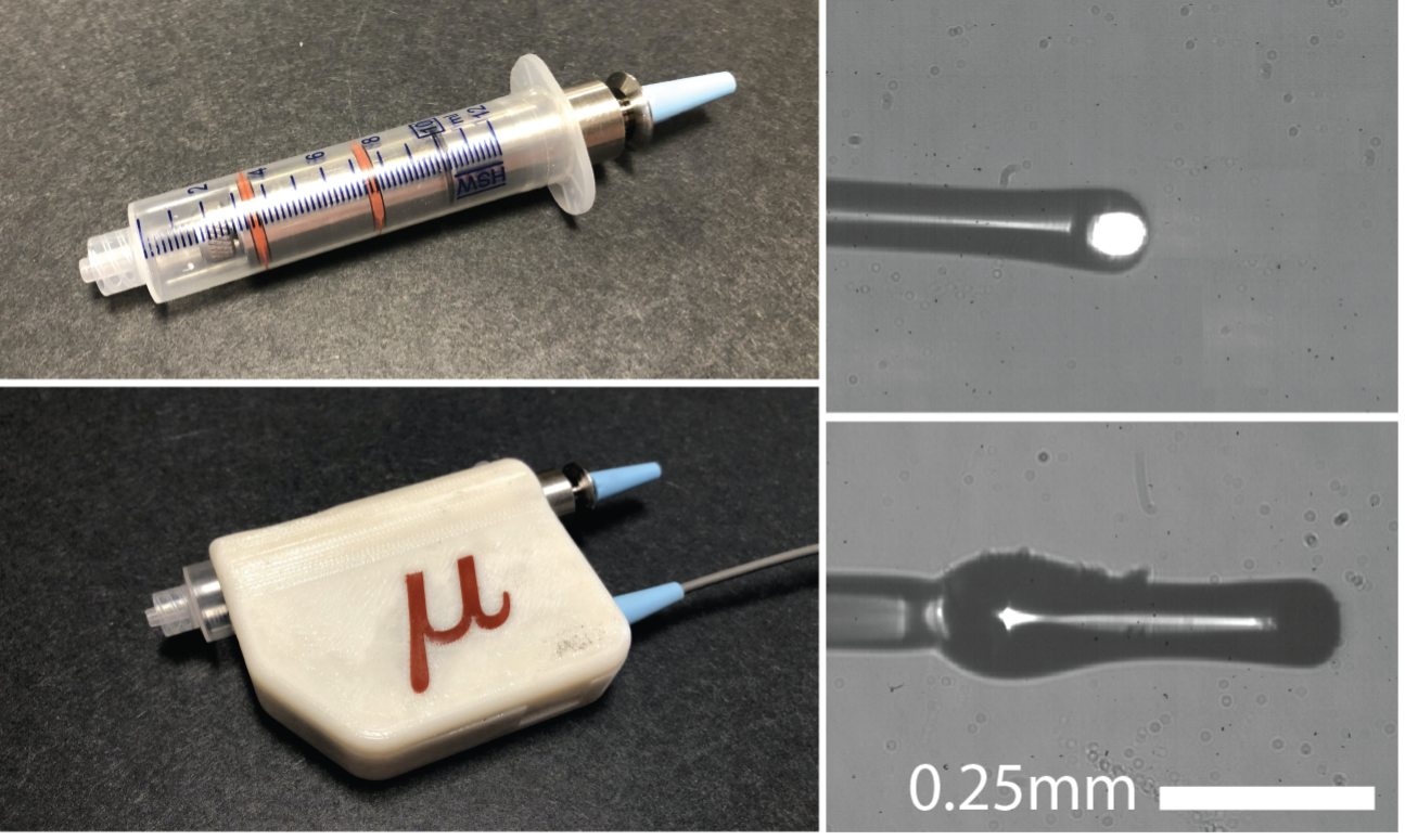 Catheter connector inside 10mL syringe and installed within the pullback unit. Photographs of angle polished ball lenses fabricated at the tip of 80µm diameter single mode fiber.
Key Researchers
Relevant Publications
- Uribe-Patarroyo, N. & Bouma, B. E. Velocity gradients in spatially resolved laser Doppler flowmetry and dynamic light scattering with confocal and coherence gating. Rev. E 94, 022604 (2016).
- Villiger, M. et al. Optic axis mapping with catheter-based polarization-sensitive optical coherence tomography. Optica 5, 1329–1337 (2018).
- Otsuka, K., Villiger, M. & Bouma, B. Intravascular Polarimetry in Patients With Coronary Artery Disease. Cardiovasc. Imaging (2019).
- Ferris, N. G., Cannon, T. M., Villiger, M., Bouma, B. E. & Uribe-Patarroyo, N. Forward multiple scattering dominates speckle decorrelation in whole-blood flowmetry using optical coherence tomography. Opt. Express 11, 1947–1966 (2020).
- Otsuka, K., Villiger, M., Nadkarni, S. K. & Bouma, B. E. Intravascular Polarimetry: Clinical Translation and Future Applications of Catheter-Based Polarization Sensitive Optical Frequency Domain Imaging. Cardiovasc. Med. 7, 1–15 (2020).